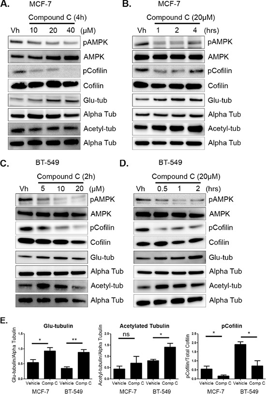
Figure 1. AMPK inhibition increases cofilin activation and microtubule stability.

A & B. MCF-7 cells were treated with 10–40 μM of compound c in serum-free media for 1–4 hours. Protein was harvested and Western blot analysis was done to determine levels of pAMPK at threonine 172, pCofilin at serine 3, detyrosinated alpha tubulin (glu-tub), and acetylated alpha tubulin (acetyl-tub). C & D. BT-549 cells were treated with 5–20 μM compound c in serum-free media for 30 mins to 2 hours. Protein was harvested and Western blot analysis was done to determine levels of markers listed above. E. Densitometry was done to quantify levels of pCofilin, glu-tubulin, and acetylated tubulin from at least three independent experiments (mean +/− SEM) in MCF-7 and BT-549 cells treated with 20 μM compound c for 4 and 2 hours, respectively. *p < 0.05. **p < 0.005. ns = not significant.
