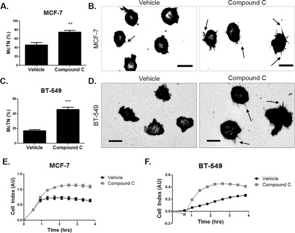
Figure 4. AMPK inhibition increases microtentacles and tumor cell re-attachment.

A. MCF-7 cells were suspended after treatment with 20 μM compound c for 4 hours in serum-free media and stained with CellMask Orange cell membrane dye. Blinded quantification of microtentacle frequency from three independent experiments (mean +/− SEM) with 100 cells counted for each was averaged. **p < 0.001. B. Representative images show microtentacles (arrow) on vehicle control and compound c treated MCF-7 cells. Scale bar corresponds to 20 μm. C. BT-549 cells were suspended after treatment with 20 μM compound c for 4 hours in serum-free media and stained with CellMask Orange cell membrane dye. Blinded quantification of microtentacle frequency from three independent experiments (mean +/− SEM) with 100 cells counted for each were averaged. ***p < 0.0001. D. Representative images show microtentacles (arrow) on vehicle control and compound c treated BT-549 cells. Scale bar corresponds to 20 μm. E & F. Re-attachment of suspended MCF-7 and BT-549 cells treated with 20 μM compound c for 4 hours and 2 hours, respectively, in serum-free media using the real time xCELLigence analyzer.
