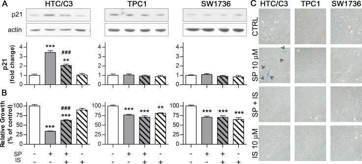
Figure 3. activated p53 induces senescence through its effector p21.
Cells were incubated with 10 μM SP either in the presence of or without p53 inhibitor Ischemin (IS, 10 μM, subtoxical concentration determined in TPC1 cells) for 96 hours followed by the assessment of p21 expression, growth inhibition, morphological changes and senescence induction. Corresponding amounts of DMSO (CTRL) or Ischemin (IS) alone were used as controls. In the panel A and B, the p21 expression and the growth inhibition data are aligned with the respective treatment scheme below the bar diagram of growth inhibition. A. Representative images of western blots and their quantification illustrating the effect of SP and Ischemin treatment on p21 expression; actin was used as loading control B. Differential effects of Ischemin on the SP-induced growth inhibition in HTC/C3, TPC1 and SW1736 cells. C. SP-induced morphological changes and senescence (detected as blue senescence-specific X-gal staining - arrowheads) are reverted by Ischemin treatment in HTC/C3 cells. **p < 0.01, ***p < 0.001 vs. control; ###p < 0.001 vs. SP treated cells.

