
Figure 2. Combined treatment with ABT263 and TIC10/ONC201 results in an enhanced induction of apoptosis.
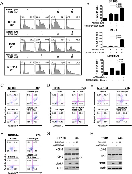
A. representative histograms of SF188, T98G and MGPP-3 glioblastoma cells that were treated as indicated with TIC10/ONC201, ABT263, both or solvent prior to staining with propidium iodide (PI) and flow cytometric analysis. B. quantitative representation of the fraction of sub-G1 cells for SF188, T98G and MGPP-3 cells treated as described for A. Columns, means of the fraction of sub-G1 cells. Bars, SD. C–F. representative histograms of SF188 (C), T98G (D), MGPP-3 (E) glioblastoma cells and NCH644 (F) glioma stem-like cells stained for annexin V/PI and treated as indicated. G–H. SF188 (G) and T98G (H) glioblastoma cells were treated for 6 h or 24 h respectively with TIC10/0NC201, ABT263 both agents or solvent under serum starvation (1.5% FBS). Whole-cell extracts were examined by Western blot for cleaved caspase 3 (cCP-3) or caspase 9 (CP-9 - CF = cleaved fragment) and cleaved PARP (cPARP). Actin Western blot analysis was performed to confirm equal protein loading.