
Figure 4. Down-regulation of Mcl-1 is mediated through enhanced proteasomal degradation.
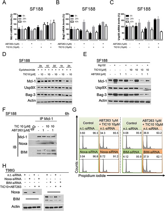
A. SF188 glioblastoma cells were treated for 6 h, 24 h or 48 h with ABT263, TIC10/ONC201, both agents or solvent under serum starvation prior to collecting RNA and performing rtPCR for MCl-1 (A), Bag3 (B) or Usp9X (C) Columns, means of the percentage of mRNA expression normalized to control. Bars, SD. D. SF188 glioblastoma cells were treated with the protein synthesis inhibitor cycloheximide (10 μg/ml) in the presence or absence of TIC10/ONC201 for the indicated lengths of time. Western blot analysis was performed for Mcl-1, Usp9X and Bag3. Actin expression was determined to confirm equal protein loading. E. SF188 pediatric glioblastoma cells were treated with ABT263, TIC10/ONC201, both or solvent in the presence or absence of the proteasome inhibitor MG132 (10 μM) for 5 h. Whole-cell extracts were collected prior to Western blot analysis for Mcl-1, Usp9X and Bag3. Actin expression was determined to confirm equal protein loading. F. SF188 glioblastoma cells were treated for 6 h with ABT263, TIC10/ONC201, both agents or solvent under serum starvation. Whole-cell extracts were collected prior to immunoprecipitation (IP) for Mcl-1. IP with murine non-specific IgG served as negative control. Western blot analysis for Mcl-1, Noxa and BIM was performed for the immunoprecipitate. G. T98G glioblastoma cells were treated with non-targeting (n.t.)-siRNA, Noxa-siRNA or BIM-siRNA followed by a treatment with TIC10/ONC201/ABT263 or solvent for 24 h. Staining for propidium iodide was performed prior to flow cytometric analysis. Representative histograms are shown. H. T98G glioblastoma cells were treated as described for G. Whole-cell extracts were collected and analysed by Western blot for Noxa and BIM to confirm successful knock-down. Actin Western blot analysis was performed to ensure equal protein loading.