
Figure 1. AF inhibits gastric cancer cells growth.
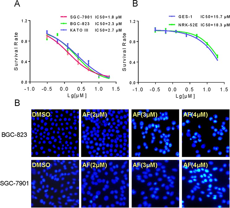
A–B. The effects of AF on the proliferation of human gastric cancer cells and normal cells. BGC-823, SGC-7901, KATO III, GES-1 or NRK-52E cells were incubated with increasing doses of AF (0.625–20 μM) for 24 h respectively. Cell viability was determined by MTT assay and the IC50 values were calculated. C. AF treatment induced apoptotic morphology in BGC-823 and SGC-7901 cells. BGC-823 or SGC-7901 cells were treated with AF (2, 3 or 4 μM) for 12 h. Cell morphology was observed using an inverted microscope after Hoechst 33258 staining.