
Figure 2. Promotion of cell proliferation by overexpression of 14-3-3γ in cancer cells.
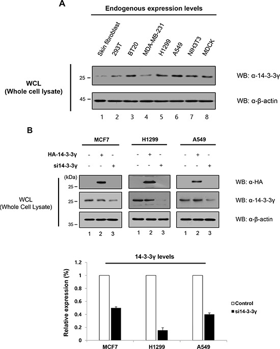
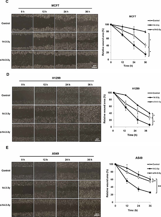
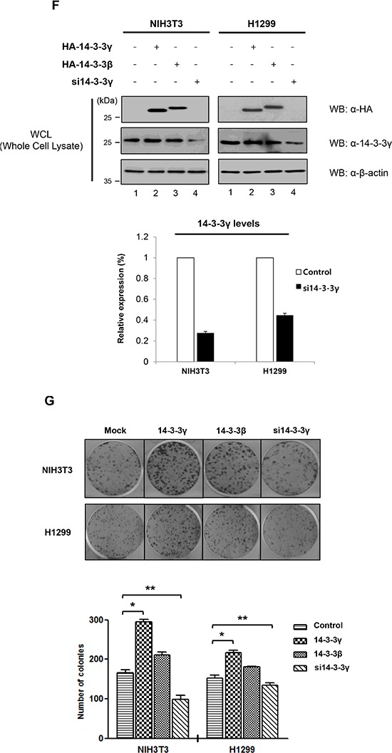
A. The expression of endogenous 14-3-3γ was investigated in various cancer cells, including breast and lung, in addition to immortalized cancerous cell lines and, normal cells. Cell lysates were used for immunoblotting with an anti-14-3-3γ antibody. B. 14-3-3γ-mediated migratory and invasive potential in MCF7, H1299, and A549 cells were investigated in a wound-healing assay. n = 3. C, D, and E. Wound healing by migrated cells at 0, 12, 24 and 36 h was imaged. Scale bar = 200 μm. The percentage of migration was statistically analyzed from separate experiments and graphed using Graph Pad Prism Software. The data are presented as means ± s.d. (Student’ t-test) *P < 0.01, n = 3. F. NIH3T3 and H1299 cells were transfected with HA-14-3-3γ and HA-14-3-3β. Additionally, siRNA specific for 14-3-3γ was used to investigate the effect of knock-down of endogenous 14-3-3γ. n = 3. H, Colony formation assay. NIH3T3 and H1299 cells stably expressing an empty vector, HA-14-3-3γ, HA-14-3-3β, and si14-3-3γ were plated in triplicate. n = 3. G. After 14 days, the colonies were stained and counted. n = 3. The number of colonies formed was graphed using Graph Pad Prism Software. The results represent the average number of colonies formed from three independent experiments. The data are presented as means ± s.d. *P < 0.01 and **P < 0.05, n = 3.