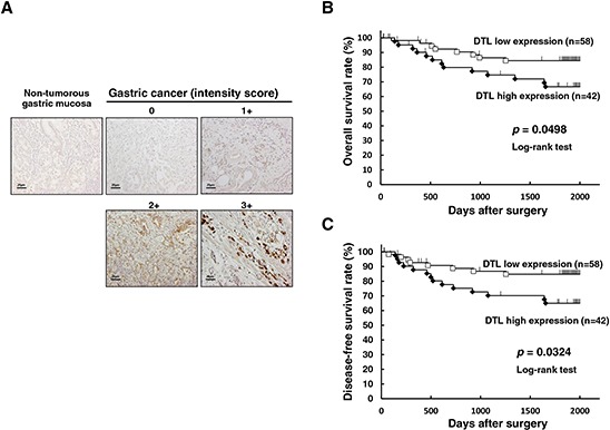
Figure 3. Immunohistochemical-staining analyses and postoperative overall survival curve according to the expression of DTL.

A. Specific immunostaining of the DTL protein in primary samples was confirmed. Expression of the DTL protein was observed in both the cytoplasm and nucleus of cancer cells. For scoring DTL expression, the intensity score was defined as 0 = negative, 1 = weak, 2= moderate, 3 = strong. The DTL high expression group had a significantly poorer prognosis than the low expression group in overall survival (P = 0.0498, log-rank test) B. and disease-free survival (P = 0.0324, log-rank test) C.
