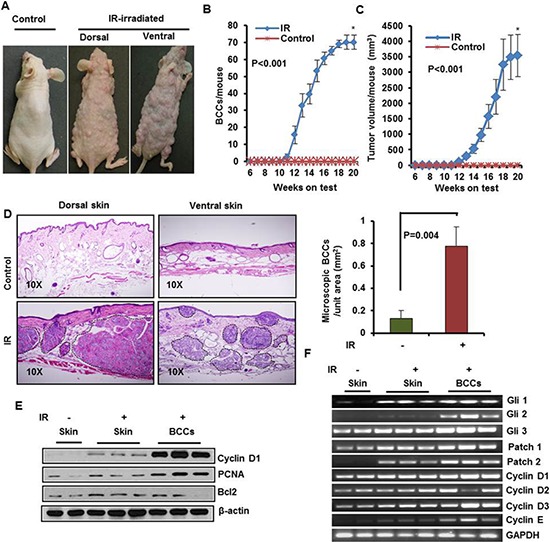
Figure 3. Ionizing radiation (IR) induce multiple BCCs in Ptch1+/−/SKH-1 mice.

A. Representative pictures of Ptch+/−/SKH-1 mice showing IR-induced visible BCCs on dorsal and ventral skin. B. BCCs/mouse. C. tumor volume/mouse; D. Histology of BCCs from dorsal and ventral skin, and analysis of microscopic BCCs/unit area (mm2) in IR-irradiated mice. E. Immunoblot analysis of biomarkers predictive of cell proliferation (PCNA and cyclin D1) and anti-apoptotic protein Bcl2 in BCCs of IR-irradiated mice. F. transcriptional expression of Glis, Ptchs and cell cycle regulatory cyclins in IR-irradiated BCC. Ptch+/−/SKH-1 mice were irradiated with a single dose (5Gy) of IR. The experiment was terminated at week 20 following irradiation. Skin and tumors were excised for histological and for molecular analysis.
