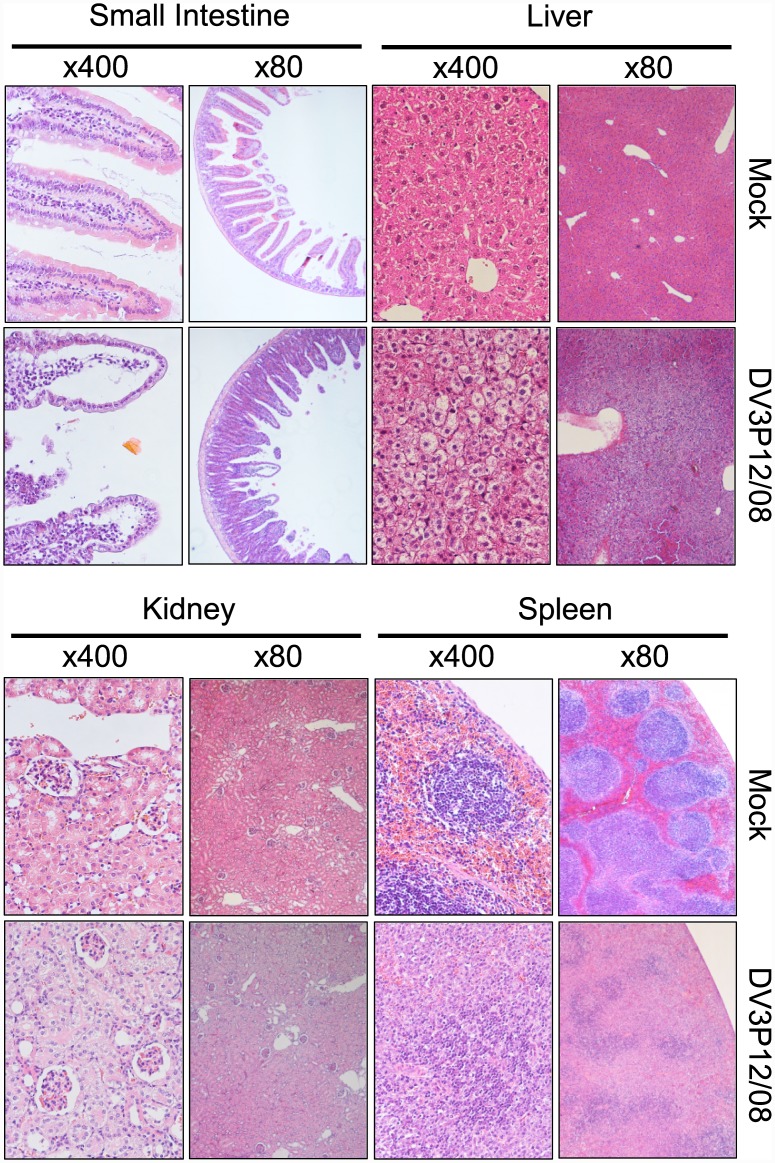
Fig 3. Histopathological examination of tissues from IFNα/β/γR KO mice infected with DV3P12/08.
IFN-α/β/γR KO mice infected with 2 × 10⁶ focus-forming units (FFU) of DV3P12/08 or mock-infected with PBS were euthanized at Day 6 p.i. Sections of liver, spleen, kidney, and small intestine were prepared, stained with hematoxylin and eosin, and observed under low (×100) and high (×400) magnification. Images are representative of at least three sections per tissue.

