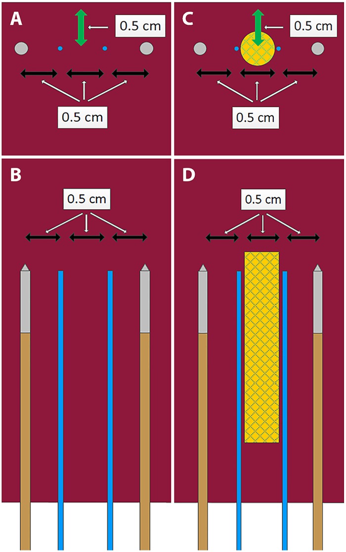
Fig 2. Setup of IRE ablations performed in in-vivo porcine liver showing the electrodes (brown/gray) and temperature probes (blue).

No-stent-IRE (A, cross-sectional; B, longitudinal) and stent-IRE (C, cross-sectional; D, longitudinal). Green arrow represents the distance to the liver surface.
