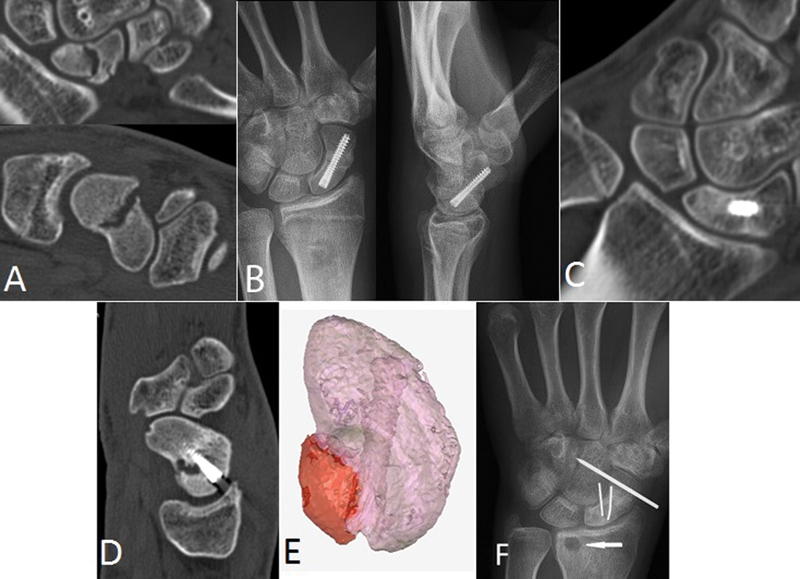
Fig. 1.

Case 1. (a) Sagittal and coronal CT images of the original mid-waist fracture nonunion. (b) Posteroanterior (PA) and lateral postoperative radiographs show central placement. (c) CT coronal view demonstrating healing at 3 months. (d) Posttraumatic CT sagittal view 4 months after healing demonstrates new fracture and cyst. (e) 3D model rendered from CT data with Mimics software (Materialise, Leuven, Belgium) shows fracture fragment (red) arising about the screw entry site. (f) Postoperative radiographs following secondary repair. A volar cortical defect from Mathoulin grafting is indicated by the arrow.
