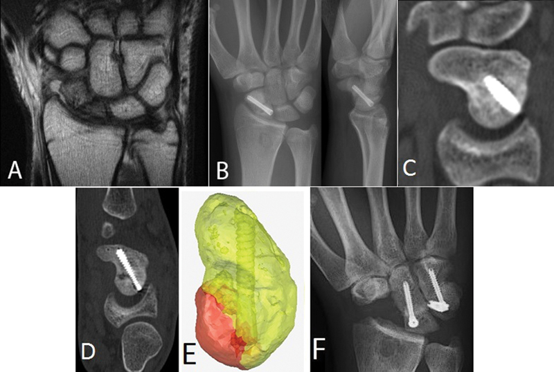
Fig. 2.

Case 2. (a) MRI 15 months postinjury demonstrates scaphoid midwaist nonunion. (b) Postoperative radiographs demonstrate central screw placement on PA and lateral views. (c) Sagittal CT view confirms healing 5 months postoperatively (d) CT 8 months after repair demonstrates de novo fracture. (e) 3D model rendered from CT data with Mimics software shows fracture fragment (red) arising about the screw entry site. (f) PA radiograph following scaphoid excision and four-corner arthrodesis.
