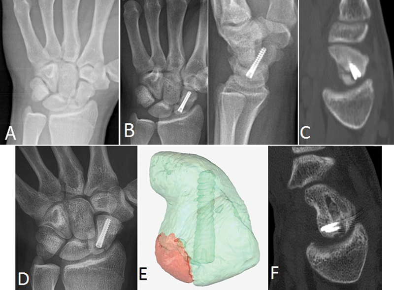
Fig. 3.

Case 3. (a) PA radiograph reveals proximal waist fracture nonunion. (b) Early postoperative PA and lateral views demonstrate good bony reduction and central screw placement. (c) Sagittal CT view showing consolidation of the fracture dorsally with incomplete healing volarly. (d) PA radiograph 18 months post repair demonstrates a lucency that resembles the original injury. (e) High-resolution CT imaging and Mimics processing reveals a fracture fragment (red) arising about the screw entry site. (f) Sagittal CT 14 weeks post secondary repair shows grafting of the original screw tract and confirms healing.
