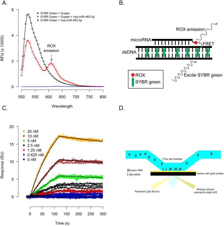
Fig 3. MicroRNAs form triplex structures with DNA.
(A) Duplex DNA identified by genome-wide screens of binding sites was incubated in presence or absence of a synthesized hsa-miR-483-5p with a 3’ ROX label to perform a FRET assay to detect triplex formation (illustrated in 3B). In the absence of ROX labeled hsa-miR-483-5p (3A, black line) a single emission peak at 520nm is observed which, with the addition of ROX labeled hsa-miR-483-5p (3A, red line), is diminished and a second FRET induced emission peak at 610nm is observed. (C) In a complementary surface plasmon resonance (SPR) based assay (illustrated in 3D), a 3’ biotin labeled hsa-miR-483-5p was immobilized and duplex DNA was introduced in triplicate in a 2-fold dilution series starting at 20 nM.

