
Figure 1.
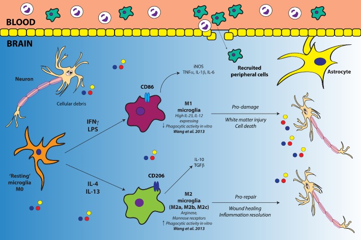
Diagrammatic representation of M1/M2 polarization in microglia. For simplicity, M2 microglia have been represented as just the one phenotype, as opposed to illustrating the various classes. Cellular debris and other mediators released by dying neurons after injury prime microglia. An environment rich with the classical pro‐inflammatory stimuli, such as IFN‐γ and LPS, promotes the polarization of resting microglia into an M1 phenotype. M1 microglia release pro‐inflammatory cytokines, chemokines and iNOS. An M1 environment is neurotoxic, facilitating white matter injury and cell death. In contrast, a neuroinflammatory environment rich in anti‐inflammatory IL‐4 or IL‐13 drives the development of an M2 phenotype. M2 microglia release IL‐10 and TGF‐β, while promoting repair and inflammation resolution. In addition to microglia, peripherally infiltrating macrophages can also undergo polarization into M1 and M2 phenotypes, and recently, this has shown to be true of neutrophils in a murine ischaemic stroke model (Cuartero et al., 2013).