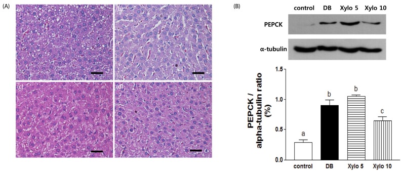
Fig. 4. Effects of D-xylose on histopathological changes in the liver, and changes in PEPCK expression in the liver.
(A) Hepatic histopathological features were observed for the four experimental rat groups: (a) normal control, (b) DB control, (c) Xylo 5, and (d) Xylo 10. (B) Levels of PEPCK in the liver were assessed using Western blotting assays. Representative blots are shown (upper panel). Band intensities were quantified by densitometry (lower panel) and were analyzed using one-way ANOVA with Newman-Keuls post hoc test (P < 0.05, n = 10 mice per group). The values shown are the mean ± SEM. Letters are used to indicate the values that significantly differ from each other (P < 0.05). Tissues were stained with H&E. Scale bar = 50 µm Magnification 400 ×. DB, diabetes control; Xylo 5 or Xylo 10, rat groups that received diets where 5% or 10%, repectively, of the total sucrose content of the diets was replaced with D-xylose; PEPCK, phosphoenol- pyruvate caroxylase.

