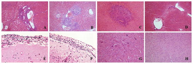
Fig. 4.
Histopathology of liver and brain tissues obtained from hamsters infected with MJNV. (A) portal inflammation in liver of MJNV-infected hamster, group I (Age<24 hr), No. 5; (B) portal inflammation and hepatic perivenulitis in liver of MJNV 04-55 infected hamster, group IV (Age 14 days), No. 8; (C) acute inflammation with necroses of hepatocytes in liver of MJNV 05-11 infected hamster, group IV (Age 14 days), No. 10; (D) liver from control uninfected hamster. (E) inflammation in brain of MJNV 04-55 infected hamster, group I (<24 hr), No. 5; (F) inflammation in brain of MJNV 04-55 infected hamster, group III (Age 10 days), No. 5; (G) acute inflammation in brain of MJNV 04-55 infected hamster, group IV, No. 8; (H) brain from control uninfected hamster. H&E stain; Original magnifications, X200.

