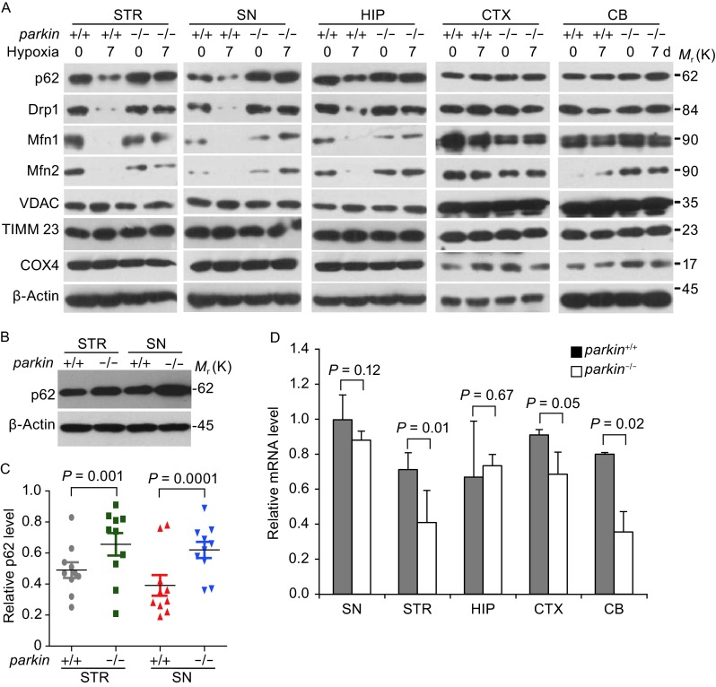
Figure 1.
P62 level is negatively correlated with parkin activity in vivo. (A) P62 is reduced in STR and SN of parkin +/+, but not parkin −/− mice under hypoxic stress. parkin +/+and parkin −/− mice, 18-month-old male C57Bl/6, were treated with 8% oxygen conditions for 0 (control) or 7 days. The striatum (STR), substantia nigra (SN), Hippocampus (HIP), frontal cortex (CTX) and cerebellum (CB) regions were isolated and homogenized in lysis buffer. Western blotting was performed to examine the level of indicated proteins. (B) P62 level increased in parkin −/− mice in STR and SN regions. The STR and SN regions from 8-week-old male C57Bl/6 mice brain were further isolated and homogenized in lysis buffer, and Western blotting was performed to examine the level of p62 (A). (C) Relative protein levels of p62 of individual mice in (1B) were quantified according to the results of ten independent blots and normalized to β-actin. (D) The mRNA levels detected by qPCR in mice tissues were described in Fig. 1B and 1D. The intensity of bands was measured with Image J software in B, mean ± SEM, from 3 independent experiments, one-way ANOVA, the P-value were indicated figures

