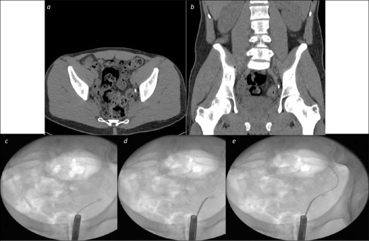
Figure 3.
Ureteric catheter to negotiate difficult stone. 3a and 3b. Axial and Coronal CT images showing a distal ureteric stone. 3c. An initial attempt to pass a wire beyond the stone failed with buckling at the site of the stone on flouroscopy 3d. A ureteric catheter over the wire, without contrast, allowed the wire to be directed towards the edge, rather than the middle of the stone 3e. The wire was passed under fluoroscopic control beyond the stone, and could then be advanced straightforwardly to the level of the kidney, and be secured as a safety guide wire.

