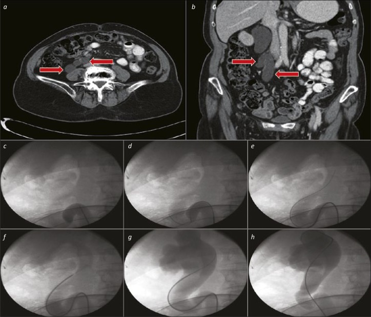
Figure 4.
Ureteric catheter for tortuous upper ureter. 4a and 4b. Axial and Coronal CT images showing a substantially dilated, tortuous proximal ureter (red arrows). 4c. The ureteric catheter is advanced as far as the beginning of the “Z” loop. 4d. A hydrophilic-tipped wire is advanced via the ureteric catheter, and onwards around the “Z” loop. 4e. The ureteric catheter and wire are negotiated upwards towards the kidney in combination. 4f. After removal of the wire, a retrograde study can be performed via the ureteric catheter to define the pelvicalyceal system anatomy (and ensure that the subsequent JJ stent is placed in the correct position). 4g. The wire is replaced via the ureteric catheter – in cases with a particularly tortuous ureter such as this, a “super-stiff” wire is often useful to aid stent placement without buckling or misplacement. 4h. The ureteric catheter is removed over the wire (and replaced with a stent – not shown).

