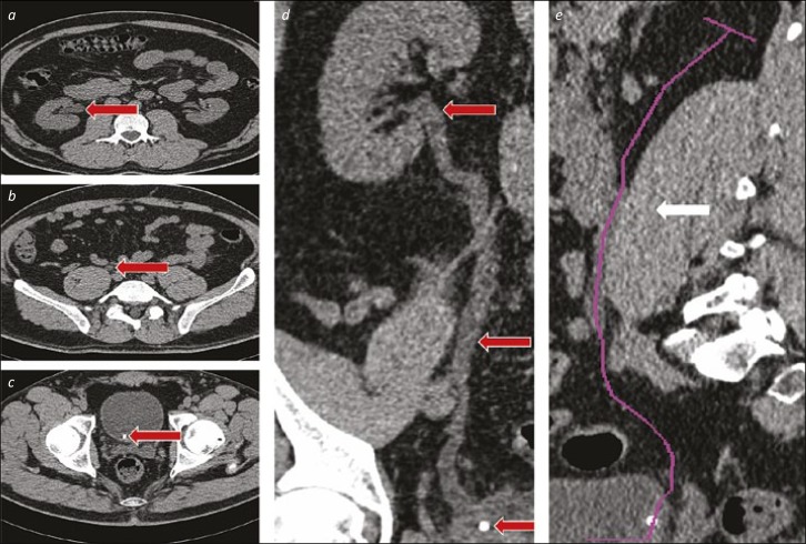
Figure 5.
The curve of the ureter. 5a, b and c. The ureter (highlighted with red arrows) is shown at the PUJ (a), in its middle third (b) and with a stone at the VUJ (c). In this series, the arrows show the movement needed from medial (at the VUJ) to lateral (at the PUJ) required for ureteroscope advancement. 5d. This full-length coronal reconstruction shows the initial course of the lower third of the ureter passes laterally, before moving medially in the middle and into the proximal ureter, before curving laterally towards the renal pelvis. 5e. The purple line on this sagittal reconstruction demonstrates the initial posterolateral direction of the ureter, and the substantial anterior displacement needed to traverse the middle third, particularly in patients with a well-developed psoas muscle (highlighted with a white arrow).

