
Fig. 9.
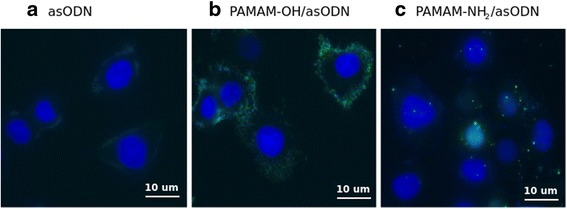
Immunofluorescence images of a free asODN, b PAMAM-OH/asODN, and c PAMAM-NH2/asODN complexes. Cells were incubated at charge ratio 1:1 in presence of dendrimers. Oligonucleotides are shown in the green channel. Cell nucleus was stained using DAPI reagent (blue)