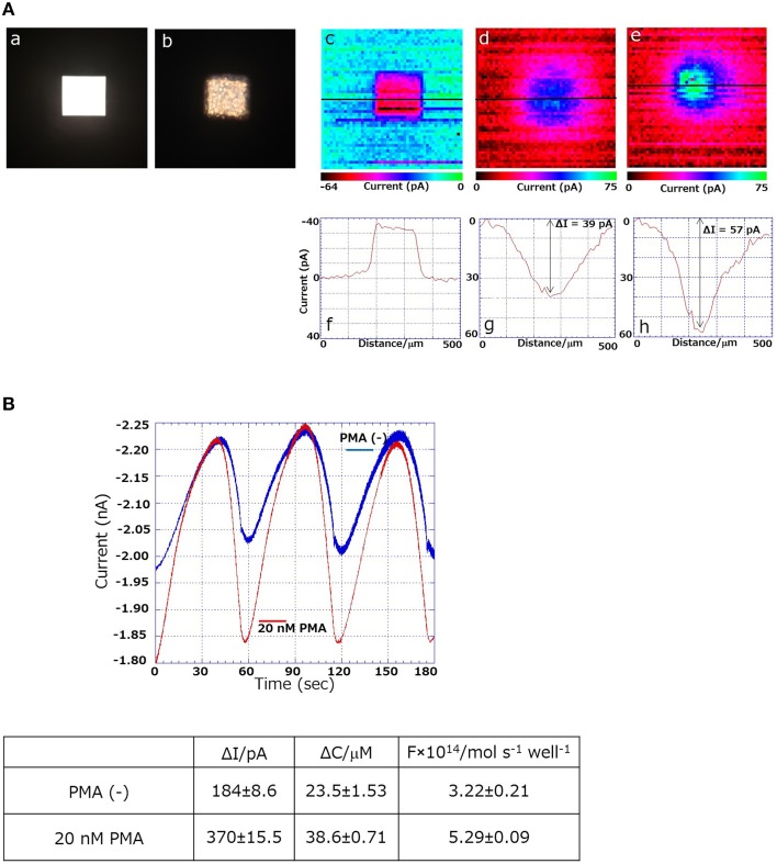
Figure 2.
(Aa) Photographs of the 3D-cell chip without (a) and with THP-1 cells (b); SECM images and changes in reduction current (in pA) using 3D THP-1 cell chip in chip containing no cells (c,f); during cellular respiration (d,g) and respiratory burst (e,h). (B) Comparison of magnitude of oxygen reduction current using SECM during ordinary respiration (blue trace) and during respiratory burst (red trace) (n = 3).

