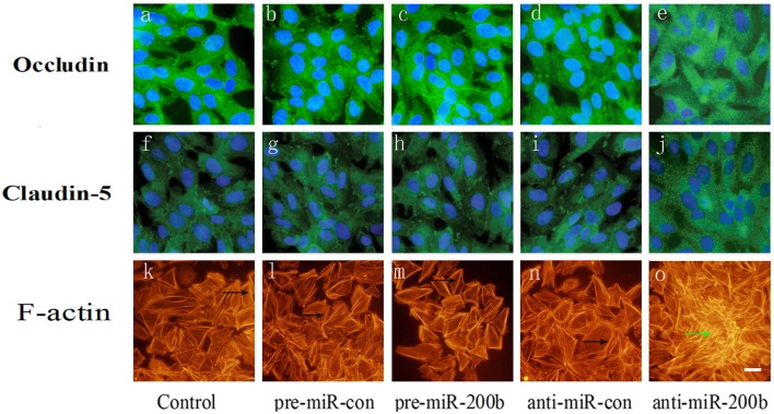
Figure 7.
Immunofluorescence analysis of occludin, claudin-5 and F-actin in GECs after overexpression or silencing of miR-200b. (A,B,D) Each represents a control group, mimic negative control group, and inhibitor negative control group in occludin. (F,G,I) Each represents a control group, mimic negative control group, and inhibitor negative control group in claudin-5. (K,L,N) Each represents a control group, mimic negative control group, and inhibitor negative control group in F-actin. Anti-miR-200b induced formation of stress fibers in the center of the cell (O) and is accompanied by a redistribution of occludin and claudin-5 from cellular membranes to the cytoplasm and nucleus (E,J). Pre-miR-200b reduced the number of central stress fibers and strongly enhanced cell peripheral localization of F-actin in GECs of the BTB (M). Furthermore, pre-miR-200b decreased the redistribution of occludin and claudin-5 from cellular membranes to the cytoplasm and nucleus and enhanced cellular membrane localization of occludin and claudin-5 in GECs of the BTB (C,H). Black arrows show the localization of peri-cellular dense aggregates of actin filaments in GECs. Green arrows show stress fibers. Scale bars represent 20 μm.

