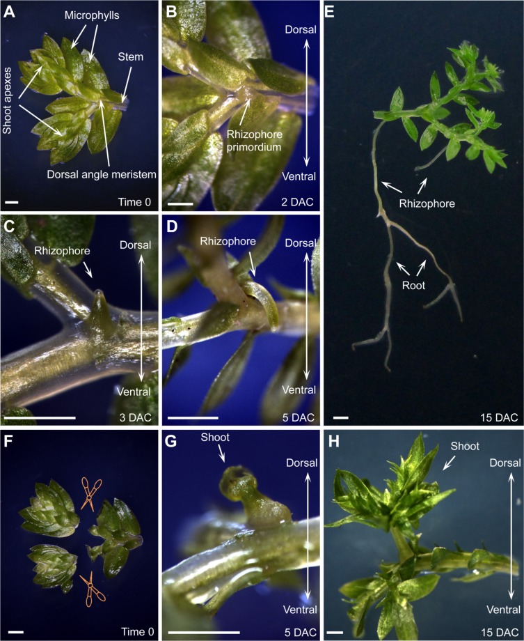
FIGURE 2.
Organ formation and regeneration in S. kraussiana. (A–D) In vitro culture of S. kraussiana, showing rhizophore growth from detached branchlets at time 0 (A), 2 DAC (B), 3 DAC (C), and 5 DAC (D). Note that rhizophore primordium was observed from the dosal angle meristem at 2 DAC (B). (E) Root formation from rhizophore. Note that the newly formed roots could bifurcate continuously. (F–H) Regeneration of shoot at angle meristem after excision of shoot apexes from branchlets. Shown are time-0 (F), 5-DAC (G), and 15-DAC (H) branchlets. Note that the regenerated shoot apex was observed from the dosal angle meristem at 5 DAC (G). Detached branchlets were cultured on wet stones and removed to an agar plate to take pictures. Scale bars, 1 mm in (A–H).

