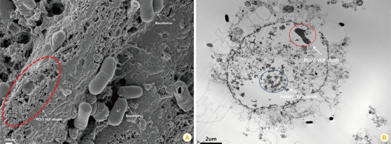
Fig. 3.
Characterization of porcine circovirus type 2 (PCV2) virus-like particles (VLPs). (A) Baculovirus and PCV2 VLPs are observed by scanning electron microscopy. Baculovirus and PCV2 VLPs were cultured in the sf9 insect cell line (scale bar=200 nm). The original size of PCV2 is approximately 15-20 nm in diameter [13]. The visible PCV2 VLPs were similar to authentic PCV2 particles in size and morphology. (B) A PCV2 VLP cluster was observed inside a sf9 cell by transmission electron microscopy (scale bar=2 µm).

