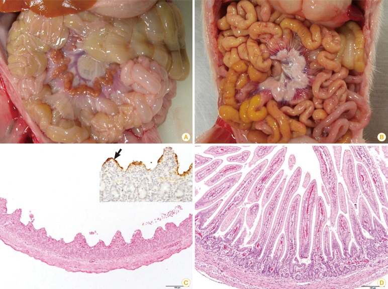
Fig. 4.
Comparison of porcine epidemic diarrhea (PED) affected (A, C) and non-porcine epidemic diarrhea virus infected (B, D) piglets. (A) The walls of the small intestine were thin and transparent. (B) No remarkable changes were found in the small intestine. (C) Severe villous atrophy and degeneration of epithelial cells were observed in the small intestine (H&E staining, scale bar=100 µm). Insert: PED antigens showed the apical portion of epithelial cells of atrophic villi (arrow, immunohistochemistry). (D) The villous height/crypt depth ratio was the range of normal sucking piglets (H&E staining, scale bar=100 µm).

