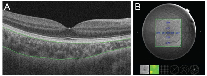
Fig. 1. Choroidal thickness map obtained using swept-source optical coherence tomography in a healthy 9-year-old girl. The 3-dimensional (D) volumetric macular scan protocol with 512 A-scans × 256 B-scans was used to obtain 3D imaging data of a 6 × 6 mm area. (A) Autosegmentation of the chorioscleral border in the B-scan image. (B) Choroidal thickness map of a 6 × 6 mm area centered on the fovea obtained from analysis of the B-scan images in the 3D data set. Mean choroidal thickness for each sector was obtained by applying the Early Treatment Diabetic Retinopathy Study grid to the map.

