. 2016 Jan 21;30(1):81–83. doi: 10.3341/kjo.2016.30.1.81

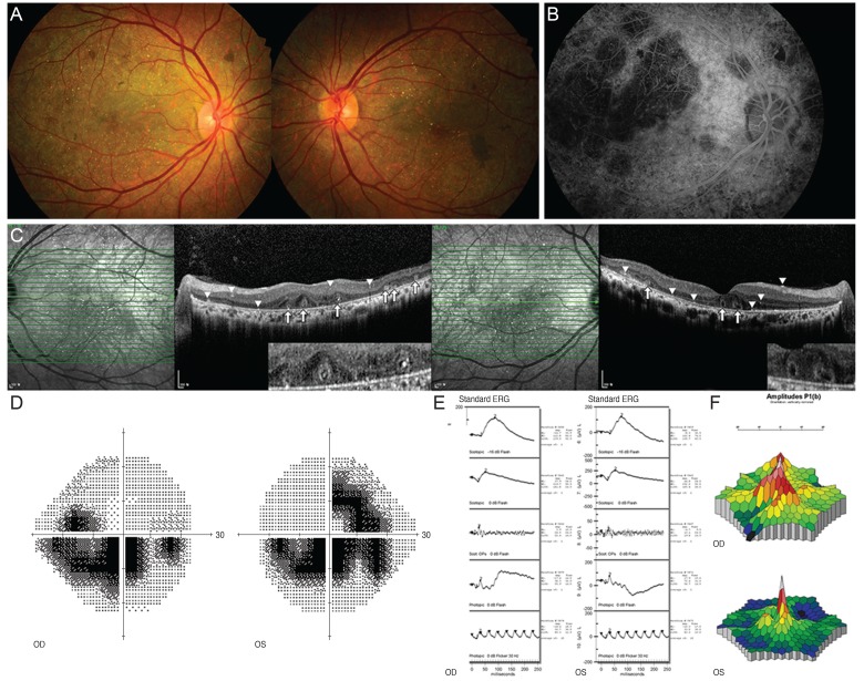
Fig. 1. Fundus photographs of both eyes (A) of our 45-year-old male patient with confirmed Bietti crystalline retinal dystrophy. Numerous retinal crystals are observed throughout the posterior pole and in the mid-periphery. Retinal pigment epithelium degeneration and choroidal atrophy are also found. A fundus fluorescence angiography image of the retinal arterial phase in the right eye (B) showing well-defined choriocapillaris filling defects, pigment epithelial window defects, and punctate blocked fluorescence. Optical coherence tomograph showing intraretinal and intrachoroidal crystalline deposits (arrowheads) with outer retinal rosette (arrows) (C). Grayscale image from Humphrey visual field test showing a central scotoma (D). Standard electroretinogram (ERG) showing slightly reduced amplitudes (E). Multifocal ERG showing reduced amplitude in the central macular area (F). OD = right eye; OS = left eye.

Fig. 1