
FIG 3 .
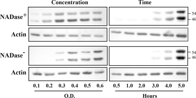
JNK activation is concentration and time dependent. Activation of JNK was assessed by Western blotting to detect its two phosphorylated forms in HeLa cell lysates following infection. The top and bottom panels contain data from cells infected with NADase+ and NADase− strains, respectively, as indicated and are compared to actin. Concentration is represented by the optical density of the infecting suspension of bacteria determined at 600 nm, and lysates for all concentration experiments were prepared at 5 h postinfection. The times shown are the hours postinfection at which cellular lysates were prepared from cells infected at an OD of 0.5. The molecular masses of proteins detected are on the right, in kilodaltons. Data are representative of at least two independent experiments.