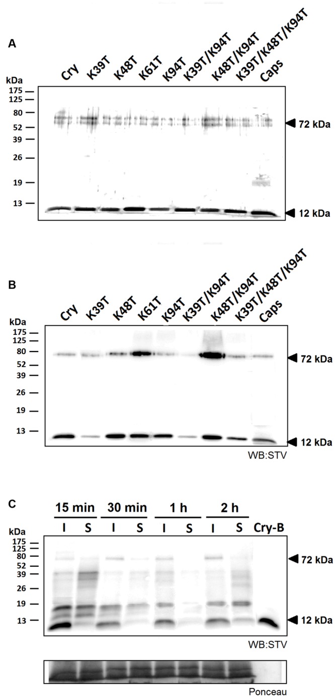
FIGURE 3.

Efficiency of biotin labeling and its effect on protein movement. The efficiency of labeling procedure was analyzed by protein detection by silver staining (A) and by western blot analysis using streptavidin labeled with horseradish peroxidase and luminescence detection (STV; B). In both cases 500 ng of proteins samples were separated and a small but visible shift in protein migration was detected as well as a significantly compromised labeling efficiency in case of proteins carrying the Lys39 mutation. Furthermore, a 72 kDa band corresponding to formed homohexamer was apparent in all tested proteins. (C) Observation of localization of biotin-labeled cryptogein in time. Samples from the site of infiltration and the surrounding tissue from three different leaves were extracted with a phosphate buffer containing 1% SDS and submitted to 15% SDS-polyacrylamide gel electrophoresis followed by western-blot analysis using streptavidin labeled with horseradish peroxidase and chemiluminescence detection (STV). In the first 30 min after infiltration a decrease in the protein concentration is visible, moreover after 2 h a small amount of protein emerged in the surrounding area. Control (CTRL) represents the samples without any treatment and Cry-B represents 5 ng of biotin-labeled wt cryptogein.
