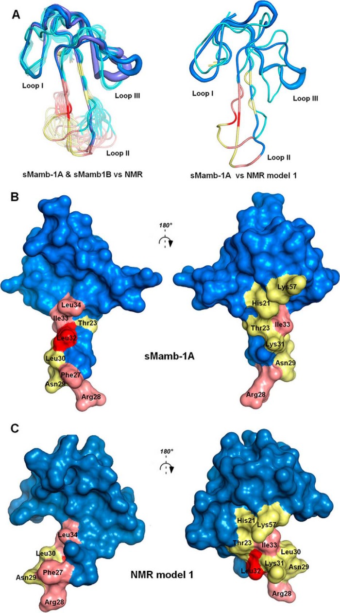
FIGURE 6.
Comparison between NMR and x-ray mambalgin structures. A, left, superimposition of all the NMR structures of sMamb-1 with the two polymorph crystallographic structures shown in blue and violet; right, profile view of the superimposition of NMR model 1 with crystal structure sMamb-1A. Visualization of the residues studied in this paper in the x-ray (B) or NMR structures (C) of Mamb-1, respectively (12, is shown. Modified residues are colored in pink for interacting and yellow for non-interacting. The key residue Leu-32 is shown in red.

