FIGURE 8.
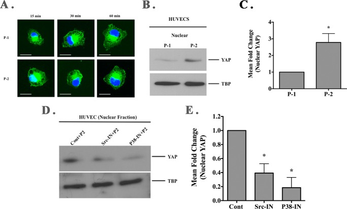
Cellular interactions with RGDKGE collagen-containing peptide facilitates nuclear YAP accumulation. A, representative examples of endothelial cells (HUVECs) attached to RGD-containing collagen peptides P-1 and P-2 stained for YAP (green). Scale bar, 50.0 μm. Photos were taken in a single plane at a magnification of ×630. B, Western blot of nuclear fraction of HUVEC lysates following binding to RGD-containing collagen peptides P-1 or P-2 probed with anti-YAP and anti-TATA-binding protein (TBP). C, quantification of the mean relative levels of nuclear YAP following ImageJ analysis. Data bars represent mean fold change ± S.E. from four independent experiments with the relative levels of nuclear YAP in cells attached to P-1 set at 1.0. D, Western blot of isolated nuclear fractions from cell lysates of endothelial cells (HUVEC) pre-incubated with Src or p38 MAPK inhibitor and stimulation with soluble RGD-containing collagen peptide P-2 and probed with YAP or TATA-binding protein (TBP). E, quantification of the mean relative levels of nuclear YAP following ImageJ analysis in cells attached to P-2 and pre-incubated with Src or p38 MAPK inhibitor. Data bars represent mean fold change ± S.E. from 2 to 3 independent experiments with the relative levels of nuclear YAP in cells under each condition with control (DMSO-treated) set at 1.0.

