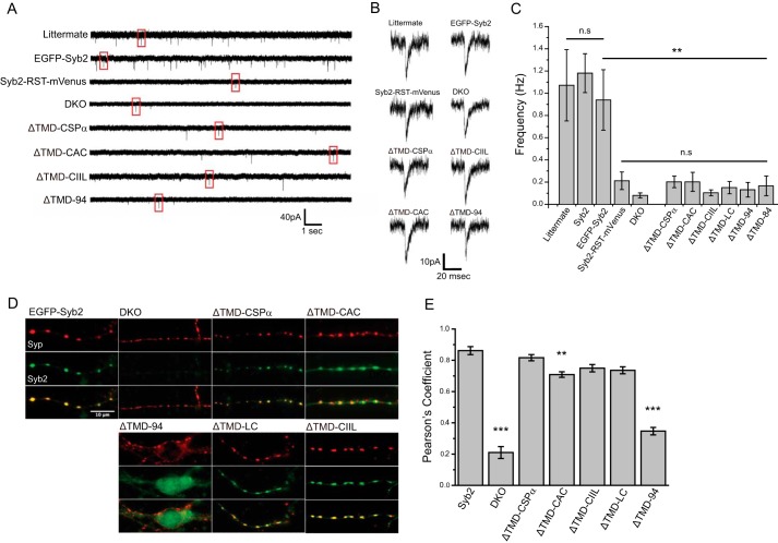
FIGURE 4.
Spontaneous synaptic release in neurons. A, patch clamp recordings of mEPSCs from littermate control and DKO hippocampal neurons, either untransfected or expressing the indicated syb2 construct. B, individual mEPSCs from each trace of A (boxed). C, mEPSC frequencies from littermate neurons and DKO neurons untransfected or expressing syb2 constructs. Syb2-RST-mVenus, lipid-anchored syb2, ΔTMD-94, and untransfected DKO cells produced similar, statistically indistinguishable low mEPSC frequencies (n = 11–30 neurons; three DKO embryos for Syb2-RST-mVenus and five for ΔTMD-LC, >9 for others). The comparison with EGFP-Syb2 or DKO is indicated by bars. D, targeting syb2 constructs in neurons. EGFP-Syb2 and lipid-anchored syb2 (green) targeted synapses labeled with synaptophysin (red). E, Pearson's coefficients indicated colocalizations of WT syb2 and lipid-anchored syb2 were well above that for ΔTMD-94, but ΔTMD-CAC was significantly lower than WT syb2. The asterisks indicate comparisons with EGFP-Syb2. **, p < 0.01; ***, p < 0.001; n.s., not significant. n = 23–44 neurons from 2 to 4 DKO embryos.

