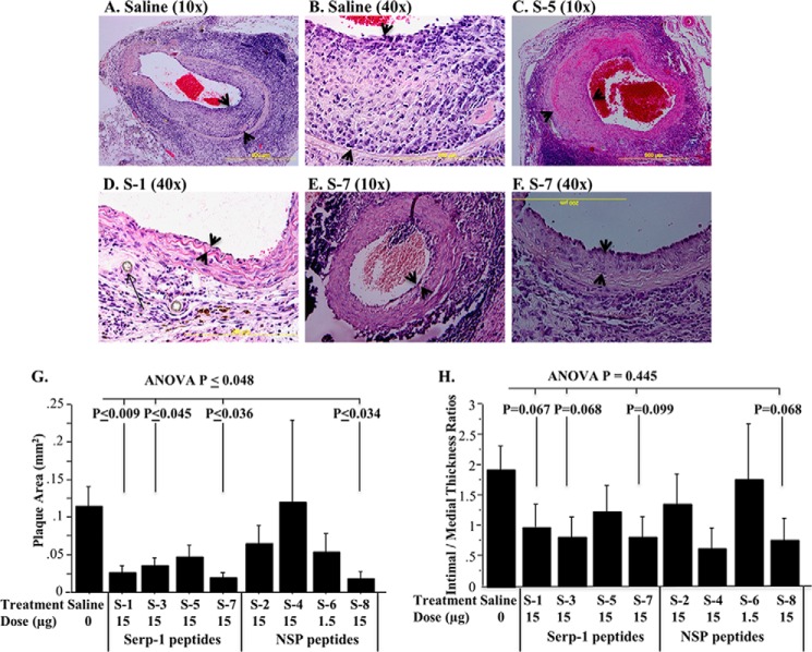
FIGURE 3.
Effects of serpin peptide infusions on plaque growth. Histological cross-sections demonstrate altered plaque growth when comparing serpin peptides (n = 93 mice, 15-μg dose immediately after transplant). Saline-treated (A, 10×; B, 40×) and S-5-treated (C, 10×) animals display larger plaque areas. S-1 significantly reduced plaque area (D, 40×), as did S-7 (E, 10×; F, 40×). The intimal hyperplasia is indicated by arrowheads. The long arrow in D shows the suture at the site of transplant. Bar graphs demonstrate significant reductions in plaque area by S-1, S-3, S-7, and S-8 peptides (G). Intimal to medial thickness ratios (H) for S-1, S-3, S-7, and S-8 also showed a trend toward reduction compared with saline controls. p ≤ 0.05 considered significant. Error bars, S.E.

