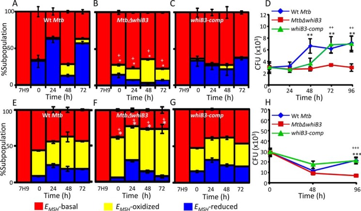
FIGURE 4.
Regulation of intramycobacterial EMSH and survival of M. tuberculosis in a WhiB3-dependent manner inside macrophages. THP-1 cells were infected with Mrx1-roGFP2 expressing WT M. tuberculosis (A), MtbΔwhiB3 (B), and whiB3-comp (C) strains at an MOI of 10. At the indicated time points, ∼30,000 infected macrophages were analyzed by flow cytometry, intramycobacterial EMSH was measured, and the percentage of bacilli in each subpopulation was determined as described earlier (15). The percentage of bacilli in each subpopulation (EMSH-oxidized, EMSH-basal, and EMSH-reduced) was plotted as a bar graph as described earlier (15). The 0 h time point refers to time immediately after infection with M. tuberculosis strains for 6 h (4 h of internalization followed by 2 h of amikacin treatment to remove any extracellular bacilli). D, THP-1 macrophages were infected with WT M. tuberculosis, MtbΔwhiB3, and whiB3-comp strains as described above, and intramacrophage survival was monitored by enumerating cfu at the indicated time points. IFN-γ- and LPS-treated (activated) RAW 264.7 macrophages were infected with Mrx1-roGFP2 expressing WT M. tuberculosis (E), MtbΔwhiB3 (F), and whiB3-comp strains (MOI of 10) (G). At the indicated time points, intramycobacterial EMSH of M. tuberculosis subpopulations was measured as described for A–C. H, activated RAW 264.7 macrophages were infected with WT M. tuberculosis, MtbΔwhiB3, and whiB3-comp strains (MOI of 2), and intramacrophage survival was monitored as described for D. The data in all panels are representative of three independent experiments performed in quadruplicate. Error bars, S.D. *, p < 0.05 (EMSH-oxidized subpopulation of MtbΔwhiB3 as compared with WT M. tuberculosis); +, p < 0.05 (EMSH-oxidized subpopulation of MtbΔwhiB3 as compared with whiB3 Comp) in experiments related to measurements of EMSH. **, p < 0.01; ***, p < 0.005 (as compared with WT M. tuberculosis); ++, p < 0.01; +++, p < 0.005 (as compared with whiB3-comp) in intramacrophage survival experiments.

