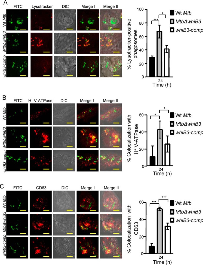
FIGURE 6.
MtbΔwhiB3 is enriched in phagosomes positive for late endosomal/lysosomal markers inside THP-1 macrophages. THP-1 cells were infected with FITC-labeled WT M. tuberculosis, MtbΔwhiB3, and whiB3-comp strains (MOI of 10). At 24 h postinfection, cells were independently stained with Lysotracker (A) or immunofluorescent antibodies for markers of phagosome maturation (i.e. V-ATPase (B) and CD63 (C), as described under “Experimental Procedures”). A representative result of three independent experiments is shown. In the images, green indicates FITC-labeled bacteria; red indicates Lysotracker or V-ATPase or CD63; differential interference contrast (DIC) indicates cell morphology; and yellow indicates merged images of two signals (Merge I, FITC/phagosomal markers) and three signals (Merge II, FITC/phagosomal markers/DIC). Scale bar, 5 μm. The bar graphs represent mean percentages of bacterium-containing phagosomes that stain positive for markers. Error bars, S.D. of three replicate wells, with each well having ∼100–200 phagosomes scored. *, p < 0.05; ***, p < 0.005 (as compared with WT M. tuberculosis); +, p < 0.05; +++, p < 0.005 (as compared with whiB3-comp).

