FIGURE 7.
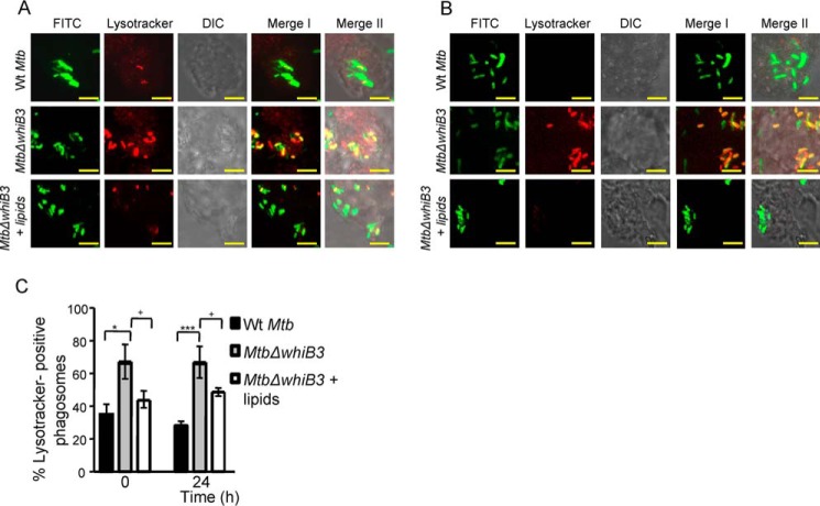
Treatment with surface-exposed lipids derived from WT M. tuberculosis reduced the enrichment of MtbΔwhiB3 in acidified phagosomes. Total surface-exposed lipids were extracted from WT M. tuberculosis and coated onto coverslips, followed by seeding of THP-1 cells as described under “Experimental Procedures.” Lipid- pretreated macrophages were infected with FITC-labeled MtbΔwhiB3 (MOI of 10). As a control, untreated THP-1 cells were infected with FITC-labeled WT M. tuberculosis and MtbΔwhiB3 (MOI of 10). At 0 h (A) and 24 h (B) postinfection, cells were stained with Lysotracker and analyzed by confocal microscopy. Representative microscopy images from at least three independent experiments for each time point are shown. In the images, green indicates FITC-labeled bacteria, red indicates Lysotracker, differential interference contrast (DIC) indicates cell morphology, and yellow indicates merged images of two signals (Merge I, FITC/phagosomal markers) and three signals (Merge II, FITC/phagosomal markers/DIC). Scale bar, 5 μm. C, mean percentages of bacterium-containing phagosomes that stain positive for Lysotracker. Error bars, S.D. of three replicate wells, with each well having >100 phagosomes scored. *, p < 0.05; ***, p < 0.005 (as compared with WT M. tuberculosis); +, p < 0.05; ++, p < 0.01 (as compared with whiB3-comp).

