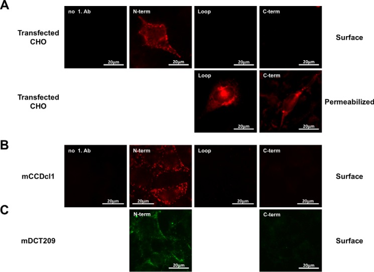
FIGURE 2.
A, surface staining of CHO-K1 cells overexpressing FLAG-tagged rLCN2-R. Live or fixed and permeabilized cells were stained with antibodies against the N terminus (α-NT-24p3R), the C terminus (PAB13044), and the loop region between the two N- and C-terminal halves of the protein (PAB20130). Fixed and permeabilized cells were also stained with an antibody against the C-terminal FLAG tag (M2) (data not shown). B and C, surface staining of cultured cortical collecting duct (mCCDcl1) (B) and murine distal convoluted (mDCT209) cells (C) expressing mLCN2-R on their surface with α-NT-24p3R (B and C), PAB13044 (B), or α-CT-24p3R (C).

