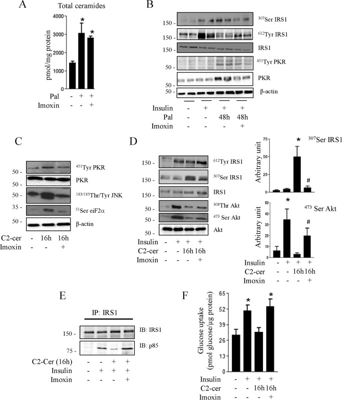
FIGURE 3.
Effect of imoxin on palmitate and ceramide-induced inhibition of insulin signaling in C2C12 myotubes. A, C2C12 myotubes were treated with 0.25 mmol/liter palmitate (Pal) with or without imoxin (1 μmol/liter) for 48 h, and the total intracellular ceramide amount was quantified. Results are mean ± S.E. (n = 3). *, p < 0.05 relative to untreated control. B, C2C12 myotubes were incubated with 0.25 mmol/liter palmitate for 48 h in the presence or absence of imoxin, followed by insulin in the last 10 min before being lysed and immunoblotted with the indicated antibodies. Representative immunoblots of at least three independent experiments are shown. C2C12 myotubes were pretreated with imoxin (1 μmol/liter) for 1 h prior to incubation with C2-ceramide (50 μmol/liter) for 16 h. C, cells were lysed, and the lysates were immunoblotted with the indicated antibodies. C2-cer, C2-ceramide. D, cells were incubated with 100 nmol/liter insulin for 10 min prior to being lysed. The histograms show densitometric quantification of Ser473 Akt and Ser307 IRS1. *, p < 0.05 relative to untreated control; #, p < 0.05 relative to cells only incubated with C2-ceramide. The representative immunoblots are from three different experiments. E, cells were lysed, and lysates were immunoprecipitated (IP) with anti-IRS1 antibody. Immunoprecipitated proteins were separated by SDS-PAGE and blotted with anti-p85 or IRS1 antibodies. The blots are representative of three separate experiments. IB, immunoblot. F, glucose transport was assessed after 30 min of 100 nm insulin incubation. Results are mean ± S.E. (n = 4). *, p < 0.05 relative to untreated control.

