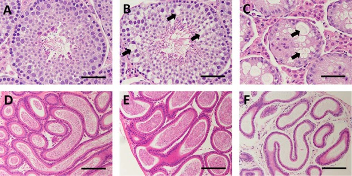
FIGURE 6.
Testicular vacuolation in Ube2w KO mice. A–C, images of H&E-stained seminiferous tubules from 16-week-old WT (A) and Ube2w KO (B and C) mice. Testes from KO mice show variable vacuolations within seminiferous tubules ranging from focal vacuolation of spermatogonia and spermatocytes (B, arrows) to diffuse vacuolation and loss of spermatocytes with marked atrophy of seminiferous tubules (C). D–F, images of H&E-stained epididymis (D–F) from the WT (D) and Ube2w KO (E and F) mice shown in A–C. Epididymis from Ube2w KO mice showed no histological abnormalities but a variable reduction in mature sperm (E and F). Scale bars, 50 (A–C) and 100 μm (D–F).

