FIGURE 8.
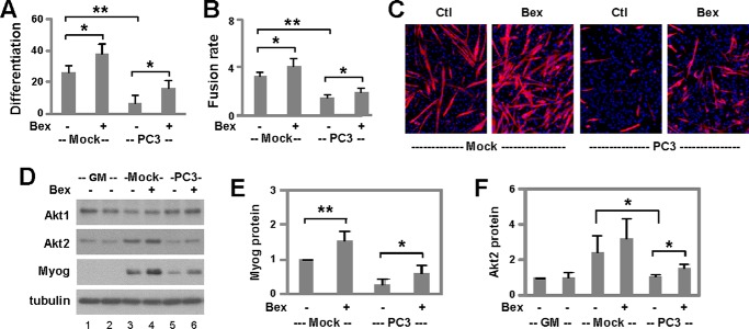
Capacity of bexarotene to retain myogenic differentiation following cachectic insult. A, C2C12 myoblasts were cultured with mock- or PC3-conditioned media for 2 days and then differentiated in fresh media in the presence of bexarotene (Bex, 50 nm) for 3 days and stained for microscopy. Differentiation was defined as the percentage of myogenic nuclei in relation to the total number of nuclei (*, p < 0.05; **, p < 0.01; n = 4). B, fusion rate was defined as the average number of nuclei per myocyte. C, representative images stained for myosin heavy chain (red) and nuclei (blue). Ctl, control. D, protein levels of Akt1, Akt2, and myogenin (Myog) on day 1 of differentiation were analyzed using Western blotting. Mock-conditioned (lane 1) and PC3-conditioned (lane 2) proliferating myoblasts (GM) were used as controls and β-tubulin as a loading control. E, quantification of myogenin protein is presented as a fold change relative to mock-conditioned untreated differentiating myoblasts (n = 5). F, quantification of Akt2 on day 2 of differentiation is presented as a fold change fold change relative to mock-conditioned proliferating myoblasts (n = 5).

