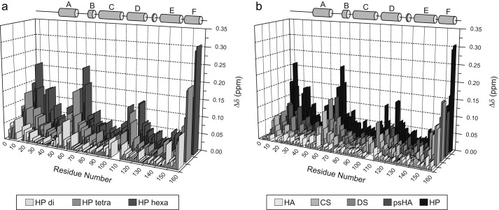
FIGURE 3.
Weighted NH chemical shift changes (Δδ) along the IL-10 primary sequence. Experiments were performed with heparins of increasing chain length (a) and different GAG oligosaccharides (b). The α-helix regions of IL-10 are indicated as gray cylinders. The protein/ligand ratio was 1:2 in each case. In b, hexasaccharides were used except for psHA tetrasaccharide. HP, heparin.

