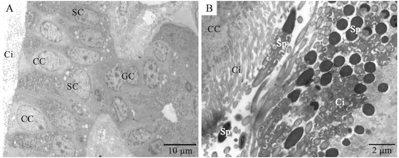
Figure 2. TEM showing the isthmus of the oviduct in P. sinensis in January.
(A) The mucosal epithelium of the oviduct comprises ciliated and secretory cells. (B) Many spermatozoa were embedded among the cilia. Secretory cell (SC), ciliated cell (CC), gland cell (GC), cilia (Ci), spermatozoa (Sp). Scale bar = 10 μm (A) and 2 μm (B).

