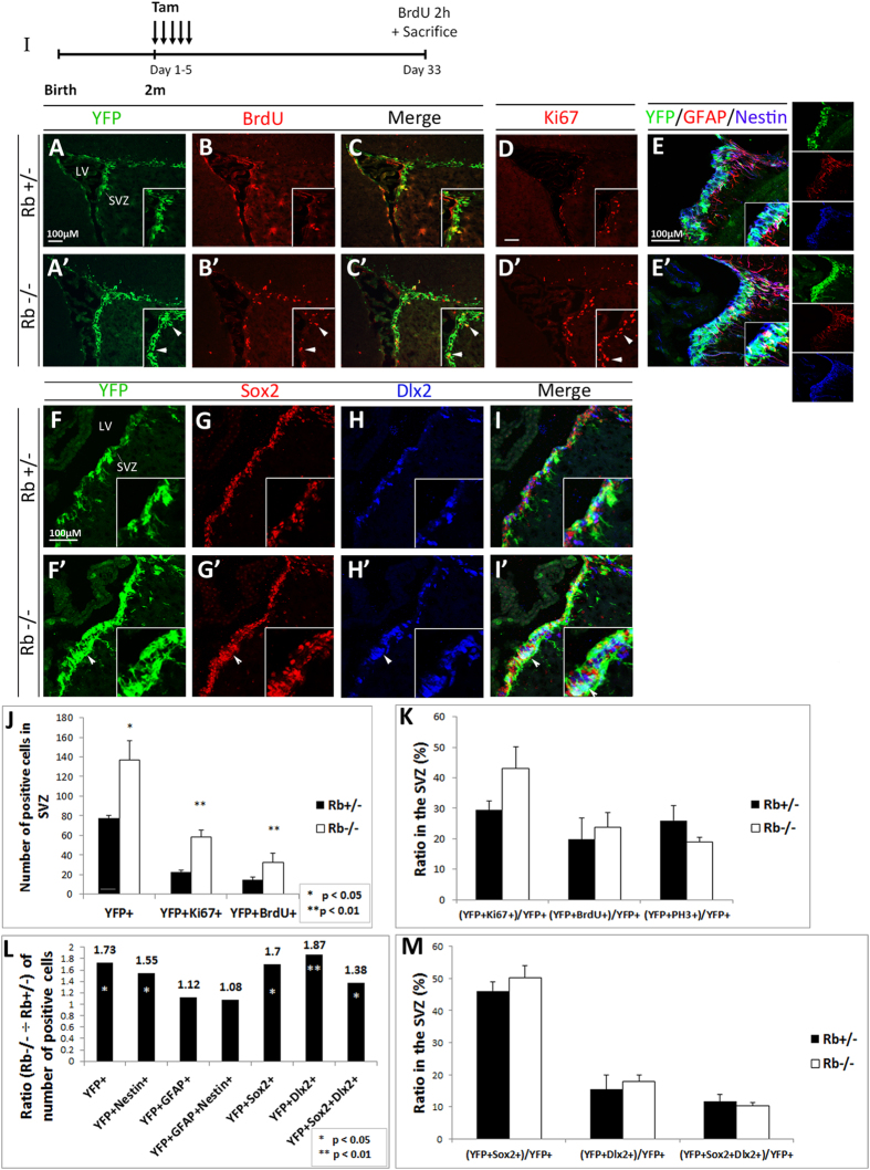
Figure 2. Rb-null mice display enhanced progenitor proliferation in the adult SVZ.
(I) Experimental design for the temporal deletion of Rb. Animals were sacrificed 28 d post-tamoxifen treatment. (A–C′) Double immunolabeling performed on sagittal sections with anti-YFP and anti-BrdU, and (D,D′) anti-Ki67 showing increased cell proliferation in the SVZ (arrowheads in insets A′–D′) (n = 5Ct and 5Mut). (E,E′) Triple staining with antibodies against YFP, GFAP and Nestin showing an increase in YFP+ and/or Nestin+ cells in the mutant SVZ compared to controls (n = 3Ct and 3Mut). (F–I′) Triple labeling with anti-YFP, anti-Sox2 and anti-Dlx2 in the SVZ in Rb+/− (F–I) and Rb−/− (F′–I′) mice highlighting the increase in distinct progenitor populations in the absence of Rb (arrowheads in F′–I′) (n = 3Ct and 3Mut). Insets in (A–I′) are higher magnifications of selected regions in the medial SVZ. (J,K) quantifications of YFP+, Ki67+ and/or BrdU+ cells in the SVZ, and the ratios of double positive cells over total YFP+ cells in Rb+/− versus Rb−/− mice, respectively. Note that the proliferation index, [(YFP+BrdU+) or (YFP+Ki67+) ÷ total YFP+], did not change between genotypes. (L) depicts the ratios (Rb−/− ÷ Rb+/−) of the different cell populations found in the SVZ; note the increase in YFP+ progenitors co-expressing Nestin, Sox2 and Dlx2. (M) graph showing the ratios of YFP-positive cells co-expressing Sox2, Dlx2 or both over total YFP+ cells in the SVZ. Error bars represent SD of measurements from at least n = 3 per genotype and asterisks indicate a statistically significant difference between genotypes using independent samples t-tests. Scale bar = 100 uM.

