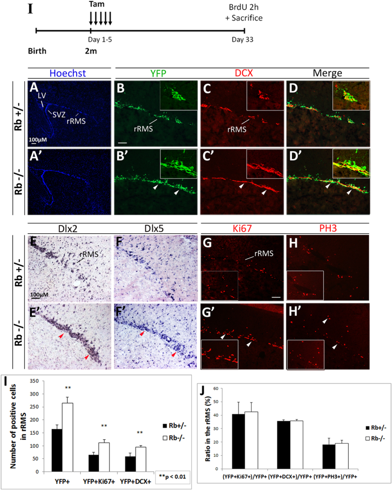
Figure 3. Rb-null neuroblasts migrate along the RMS to the OB similar to Rb+/− control cells.
(I) Experimental design for the temporal deletion of Rb. (A–D′) Immunolabeling with Hoechst, YFP and DCX on sagittal sections showing an increase in the number of neuroblasts migrating in the rostral part of RMS (rRMS) in Rb−/− versus Rb+/− (arrowheads in B′–D′) (n = 3Ct and 3Mut). This increase was confirmed by in situ hybridization using RNA labeled anti-sense probes for two neuronal differentiation markers; Dlx2 (E,E′) and Dlx5 (F,F′) (red arrowheads in E′,F′) (n = 3Ct and 3Mut). The enhanced neuroblast migration was associated with higher proliferation in the rRMS and normal cell cycle exit as shown by Ki67 (G,G′; arrowheads) and PH3 staining (H,H′; arrowheads), respectively. (I) Quantification of YFP+ cells co-labeled with DCX or Ki67 in the rRMS. (J) Graph showing the proliferation, migration and mitotic indices [double positive cells ÷ total YFP cells] in both genotypes. Error bars represent SD of measurements from n = 3 per genotype and asterisks indicate a statistically significant difference between genotypes using independent samples t-tests. Scale bar = 100uM.

