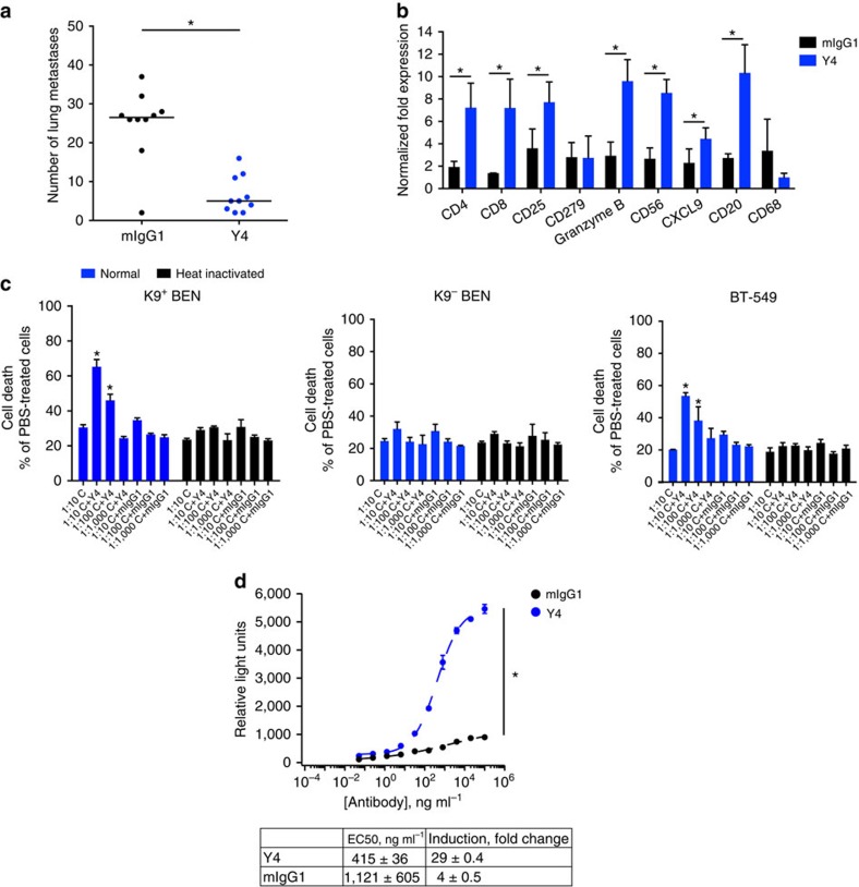
Figure 6. Y4 inhibits lung metastasis and induces immune responses.
(a) Number of lung metastasis foci in BALBc/ByJ mice treated with mIgG1 or Y4 i.p. twice a week for 25 days. The centre line represents median, n=10 per group, *P<0.01, two-tailed Student's t-test. (b) Expression of immune markers verified by quantitative reverse transcriptase–PCR (qRT–PCR) of mRNAs extracted from lung tissues. Mean±s.d., n=5, *P<0.05, two-tailed Student's t-test. (c) Y4 activates complement, resulting in complement-dependent cytotoxicity in vitro. K9+ BEN cells and BT-549 cells were treated with Y4 for 2 h in the presence of either normal or heat-inactivated murine complement before LDH measurement. Mean±s.d., n=6, *P<0.05, two-tailed Student's t-test. (d) Y4 activates ADCC reporter cells. BEN cells were incubated with different doses of Y4 and mIgG1, and engineered murine Jurkat T cells stably expressing FCγRIII and an NFAT-response element that drives luciferase expression (NFAT-RE-luc2). Measured luciferase activity indicates ADCC induction. Mean±s.d., n=3, *P<0.01, two-tailed Student's t-test.

