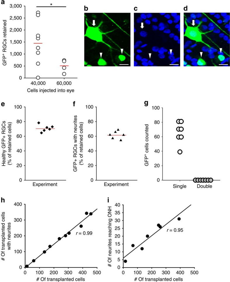
Figure 2. Transplanted GFP+ RGCs survive and extend neurites in the host retina.
(a) Retention of donor GFP+ RGCs 3–4 weeks after successful intravitreal transplantations, with significantly different mean values shown as red lines (*P<0.05 by t-test, two-sided). (b–d) Representative images from recipient, DAPI-stained retinal whole mounts showing nucleus morphologies (blue) and the corresponding green GFP-merged images of donor RGCs; examples of punctate, unhealthy nuclei (arrowheads) and large, circular, healthy nuclei (arrow) are marked. (e) Based on DAPI morphology, 70% of the retained GFP+ RGCs were judged to be healthy, and (f) over 60% extended axon-like neurites in the host retina. (g) Of nearly 500 GFP+ RGCs counted, all had single nuclei and none with double or fused nuclei were observed in the host retinas (n=7). (h,i) For GFP+ cells, the fraction of cells with neurites was constant (r=0.99), independent of the number of cells transplanted; the number of neurites reaching the optic nerve head (ONH) within the same quadrant increased linearly with cell number (correlation coefficient r=0.95), indicating that neurite growth too was not influenced by transplanted cell density. Scale bars, 20 μm.

