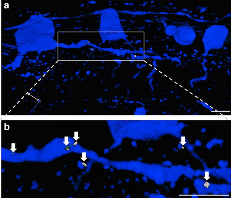
Figure 4. Transplanted RGCs form multiple synapses within the host retina.
(a) Section from recipient host retina 4 weeks post transplantation showing GFP+ transplanted RGCs in blue and synaptic puncta in white. (b) Magnified view of a portion of a GFP+ dendrite with visible synaptic puncta (white, arrows). Scale bar, 25 μm.

